Overview of Tembusu Virus
Tembusu disease is an infectious illness caused by the Tembusu virus, part of the Ntaya virus group in the Flavivirus genus. It causes laying ducks’ egg production to plummet (from 90% to 10% or even stop altogether), reduces appetite in ducklings, and slows their growth. Originally called “duck hemorrhagic oophoritis” or “duck viral encephalitis,” the virus was first found in meat ducks in 2011. By 2012, it was confirmed as the duck Tembusu virus. So far, it can infect waterfowl, commercial laying hens, and breeder chickens.

Pathogenic Characteristics of Tembusu Virus
Classification Standards
Tembusu disease belongs to the Flaviviridae family, Flavivirus genus, Ntaya virus group.
Morphological Features
The virus particles are spherical, 45–50 nm in size, and mature particles are wrapped in a phospholipid bilayer featuring glycoprotein spikes and an icosahedral nucleocapsid.
Genomic Structure
The DTMUV genome is a single-stranded positive-sense RNA virus, about 11 kb long with 10,990 nucleotides. It encodes three structural proteins: capsid protein (C), prM protein (precursor to M protein), and envelope protein (E); and seven non-structural proteins: NS1, NS2A, NS2B, NS3, NS4A, NS4B, and NS5. The C protein has low amino acid similarity, contains many basic amino acids, and helps in virus assembly. The prM gene produces the prM protein, which matures into the M (membrane protein). The M protein binds tightly with C and E proteins and aids in virus packaging.
Pathogenic Features
The virus particles are spherical, 45–50 nm in size, with mature particles enclosed in a phospholipid bilayer with glycoprotein spikes and a nucleocapsid showing icosahedral symmetry.
- DTMUV is sensitive to ether, chloroform, and deoxycholate. It’s heat-sensitive, losing activity after 60 minutes above 50°C.
- DTMUV can grow in duck embryos, chicken embryos, duck embryo fibroblasts, and Vero cells but not in chicken embryo fibroblasts.
- DTMUV mainly replicates in the cytoplasm of infected cells. When injected into the allantoic cavity, duck embryos die within 3–5 days, showing thickened and swollen chorioallantoic membranes, general edema, bleeding, and enlarged, mottled, necrotic livers.
Epidemiological Features
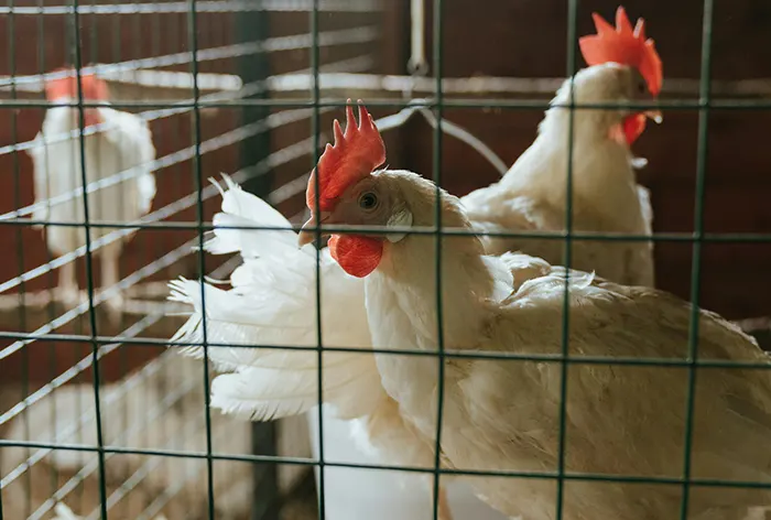
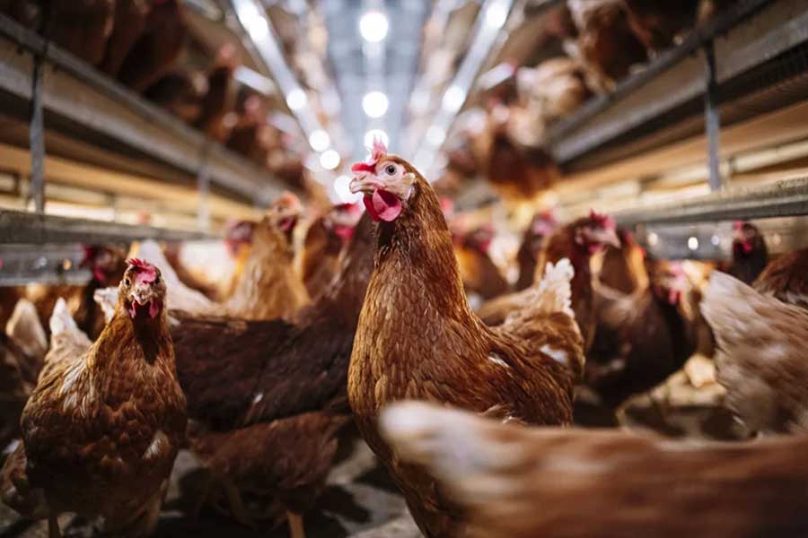
Susceptible Animals: The disease can infect waterfowl, commercial laying hens, and breeder chickens.
Transmission Routes: It spreads horizontally and vertically, with wild birds and mosquitoes playing significant roles. Naturally, the Flavivirus genus members are spread mainly by arthropods. Of over 70 Flavivirus members, 30 are mosquito-borne, and 11 are tick-borne. The disease can occur year-round, with the worst outbreaks in fall and winter. Birds are also key in its spread. The virus can be isolated from the cloaca, showing it spreads through feces, contaminating the environment, feed, water, tools, and transport vehicles.
Epidemic Characteristics: The disease has no clear seasonal pattern and can occur anytime, with laying ducks hit harder in winter and ducklings in summer.
Pathogenesis
Once inside the body, the Tembusu virus mainly attacks vascular endothelial cells, causing viremia and then affecting the spleen, reproductive organs, and brain, with the spleen likely hit first. Early on, the virus is most detectable in blood; later, it’s found in the spleen, ovarian follicles, and brain. It’s also present in the throat and feces of sick ducks. Massive viral replication and cell breakdown products flood the bloodstream, triggering inflammation and toxemia. This leads to body-wide metabolic issues (less eating and egg-laying), fever, organ dysfunction, and structural damage. It can cause necrotic and proliferative splenitis (spleen shrinks early, then swells and darkens), interstitial hepatitis and nephritis, and viral encephalitis.
Harmfulness
The Duck Tembusu virus is highly dangerous to ducklings under 7 weeks, especially those under 2 weeks, with infection and illness rates above 90% and mortality rates of 5%–30%. Ducks aged 2–4 weeks have mortality rates up to 40%, while 5–6-week-olds reach 25%. In growing ducks, harm depends on age: 7–21-week-olds are at risk, especially 7–10 and 18–21 weeks, while 14–16-week-olds resist better. Mortality in 7–8-week-olds is below 10%. Daily egg production may drop 5%–20% in laying ducks, with mortality around 2%–5%. The disease spreads fast, often infecting a whole flock in 2 days. It can also spread vertically, lowering hatching rates, increasing dead embryos, and causing weak ducklings.
TMUV Virulence Analysis
Isolates from 2012 showed no significant genetic differences from 2010–2011. In September 2019, an isolate from Jinyun, Zhejiang, in Ma ducks had a clinical mortality rate of about 60%. Confirmed as Tembusu virus via PCR, it was named JS201909. SPF chicken embryo tests showed an ELD50 of 10⁻⁵.⁴³, more potent than the traditional virulent strain ZJ407 (ELD50 ~10⁻³). Genetic analysis showed differences from earlier duck-derived Tembusu isolates, with closer ties to recent southern China isolates and striking similarity to Thai isolates, calling for tighter monitoring to prevent cross-border spread. Since 2023, NS5 gene analysis of 22 Tembusu virus strains showed homology above 98%.
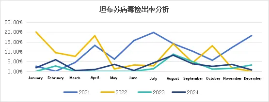
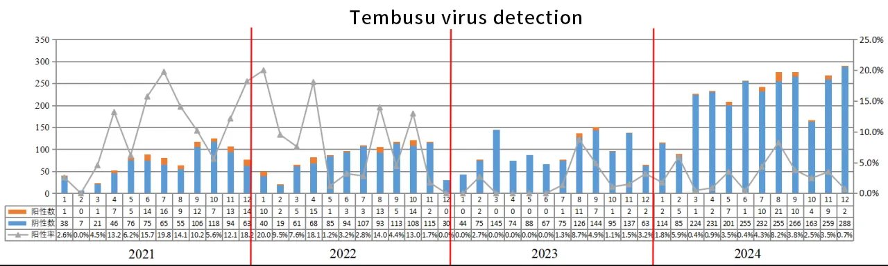
Current Status of Tembusu Virus Epidemic
Detection Rates Since 2021, Tembusu virus positivity rates have dropped thanks to widespread vaccine use. In 2024, of 2,647 samples tested, 2,573 were negative, and 74 were positive, giving a positivity rate of 2.8%. Waterfowl faced high disease rates in February, April, August, and November 2024, needing extra caution.
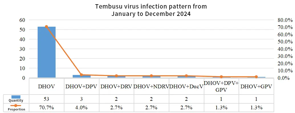
Infection Patterns In 2024, Tembusu virus infections were mainly single, with 17 types of mixed infections noted.
Clinical Manifestations
Clinical signs in ducks vary by infection stage:
Within 1 week: High mortality in ducklings, with infections causing ~20% deaths.
3–5 weeks: Culling rates of 5%–10%, up to 30% in severe cases, with secondary infections pushing mortality to 60%; stunted growth in meat ducks.
Laying period: Ducks eat 30%–60% less, egg production crashes by 10%–90%, mortality is low at 5%–15%, with an average loss of 15–20 eggs per duck, costing 45–60 yuan per duck.
Breeder ducks/geese: Fertilization and hatching rates of eggs drop by ~10% during infection.

Clinical Signs
The sharp drop in egg production: Laying ducks produce far fewer eggs, losing an average of 15–20 good eggs daily.
Stunted growth in ducklings: Ducklings infected at 1 day old gain 10%–20% less weight by 11 days.
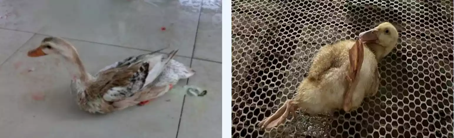
Soft legs and digestive issues: Sick ducks have weak legs and pass green feces.
Neurological symptoms: Infected ducks and geese show paralysis and lack of coordination.
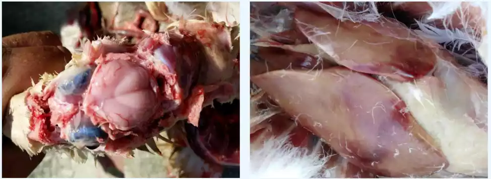
Necropsy Features of Duck Tembusu
Ovarian Lesions: Follicular bleeding. Follicular liquefaction.
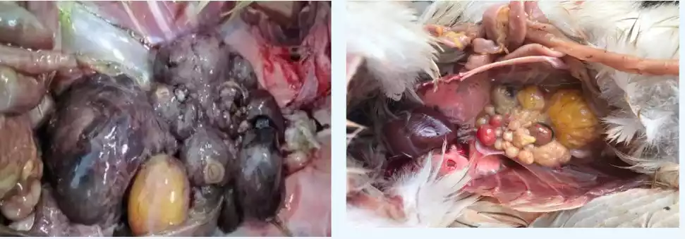
Brain Lesions: Branch-like bleeding. Brain tissue swelling.
Liver Lesions: Swollen liver with bleeding and a thinned surface showing mottled necrosis.
Heart Lesions:
- Thickened heart muscle.
- Yellow fluid at the heart base.
- Damage in the heart’s inner lining.
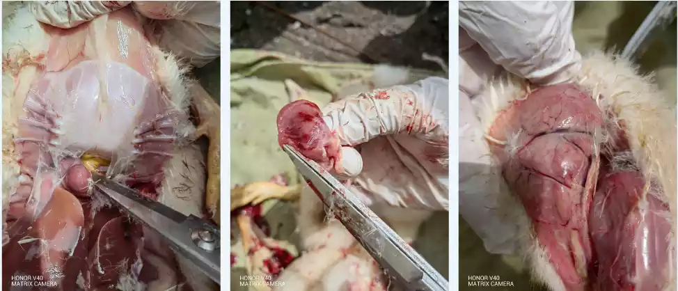
Other: Thymus shrinkage.
Control Measures
Biosecurity Control Using standard disease control methods tailored to the Tembusu virus, set up practical biosecurity steps.
Selection of High-Quality Breeder Stock
Choose breeder ducks with potent maternal antibodies (e.g., vaccinated with “one live, two inactivated” or “three inactivated” vaccines) to shield ducklings from early wild virus infections.
Maternal Antibody Decline Patterns
South: Full protection before 10 days, negative by 15 days.
North: Negative by 18 days.
Blocking Transmission Routes
Environmental Hygiene:
- Set up a solid disinfection routine, regularly cleaning farms and nearby areas.
- Clear sewage and trash, removing hygiene dead zones.
Mosquito and Fly Control: Cut down mosquito breeding and wipe out mosquitoes and flies on duck farms (leading virus carriers).
Equipment and Egg Management:
- Strictly disinfect tools, equipment, transport vehicles, and hatching eggs.
- Quickly burn or biologically process dead ducks; immediately disinfect contaminated areas and tools.
Stress Reduction: Watch weather changes during vaccination, using cold-proof and insulation measures.
Disinfection Measures: The virus is sensitive to acids, ether, deoxycholate, trypsin, and high heat; pick disinfectants suited for farms.
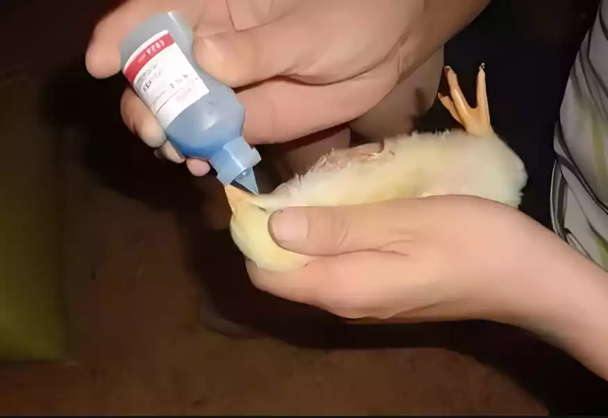
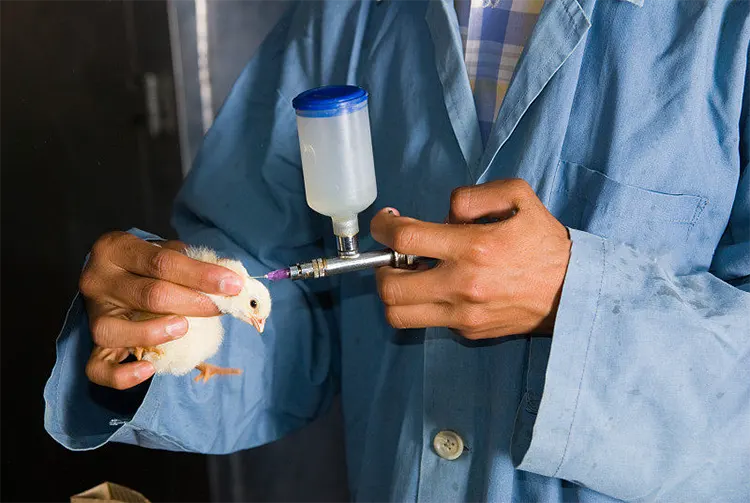
Vaccination Protocols
Recommended vaccination schedules by duck type and age:
Meat Ducks, Ma Ducks, Muscovy Ducks
Slaughter >40 days: 5–7 days, Tembushu vaccine (0.3 ml/duck).
Slaughter <40 days: 5–7 days, Yahuangping vaccine (1 dose).
Laying Ducks
5–7 days: Tembushu vaccine (0.3 ml/duck; in high-risk areas, add Yahuangping vaccine, one dose).
28 days: Tembushu vaccine (0.5 ml/duck).
2 weeks pre-laying: Tembushu vaccine (0.8 ml/duck).
Breeder Ducks
7–12 days: Tembushu vaccine (0.3 ml/duck; in high-risk areas, add Yahuangping vaccine, one dose).
28–33 days: Tembushu vaccine (0.5 ml/duck).
2 weeks pre-laying: Tembushu vaccine (1.0 ml/duck).
Commercial Geese
1 day: Yahuangping vaccine (1.2 doses).
Breeder Geese
10 days: Yahuangping vaccine (1.5 doses).
50 days: Tembushu vaccine (0.8 ml/goose).
2 weeks pre-laying: Tembushu vaccine (1.0 ml/goose).
Enhancing Immunity
Medication Support
5–7 days: Use Fanyiping + Kangtiyou (1 set per 1,000 ducks, 2 days) to boost antiviral defenses.
16–20 days (immune gap):
Kangtiyou: 1 bottle/1,000 ducks, 3 days.
Kanglineng: 1 bag/2,000 kg water, 5 days.
Benefits: Eases immune suppression, strengthens antiviral defenses, and boosts overall flock resistance.
Benchmark Case Study
Guangxi Group
Used avian influenza vaccine + Tembusu inactivated vaccine + ceftiofur (Beifuxin) for combined immunization.
Results
21 days post-immunization: Antibody level 6log2, Flavivirus antibody positivity rate 93.3%.
31 days post-immunization: Antibody level 7log2, positivity rate 100%.
After 20 days: Lower culling rates, better leg strength, higher survival, better feed-to-meat ratio, increased weight, and more significant profits.
Promotion: Rolled out fully from September 2021, topping breeding results 2022.
Control Priorities
High-Risk Periods: Boost monitoring and vaccination in February, April, August, and November.
High-Risk Areas: Anhui, Guangdong, and Jiangxi need stronger biosecurity and immunization.
Virus Mutation: Watch for strain changes (e.g., more substantial 2019 JS201909 strain) and ramp up testing to stop cross-border spread.